Gold Injected Into Your Cells Gold Used as Art

Suspensions of gold nanoparticles of various sizes. The size difference causes the difference in colors.
Colloidal gilded is a sol or colloidal suspension of nanoparticles of gold in a fluid, unremarkably water.[1] The colloid is usually either an intense red colour (for spherical particles less than 100 nm) or blue/purple (for larger spherical particles or nanorods).[2] Due to their optical,[3] electronic, and molecular-recognition properties, gold nanoparticles are the subject area of substantial inquiry, with many potential or promised applications in a wide variety of areas, including electron microscopy, electronics,[4] nanotechnology, materials science,[five] and biomedicine.[vi] [7] [viii] [9]
The properties of colloidal aureate nanoparticles, and thus their potential applications, depend strongly upon their size and shape.[10] For case, rodlike particles have both a transverse and longitudinal assimilation peak, and anisotropy of the shape affects their self-assembly.[11]
History [edit]
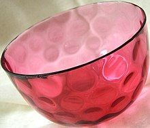
This cranberry glass bowl was made by adding a aureate salt (probably gold chloride) to molten glass.
Used since ancient times as a method of staining glass colloidal gold was used in the quaternary-century Lycurgus Cup, which changes colour depending on the location of light source.[12] [13]
During the Center Ages, soluble gold, a solution containing gold salt, had a reputation for its curative belongings for diverse diseases. In 1618, Francis Anthony, a philosopher and member of the medical profession, published a volume called Panacea Aurea, sive tractatus duo de ipsius Auro Potabili [fourteen] (Latin: aureate potion, or ii treatments of potable gold). The volume introduces information on the germination of colloidal gold and its medical uses. Well-nigh half a century later on, English botanist Nicholas Culpepper published book in 1656, Treatise of Aurum Potabile,[15] solely discussing the medical uses of colloidal gold.
In 1676, Johann Kunckel, a High german chemist, published a book on the industry of stained drinking glass. In his volume Valuable Observations or Remarks Nearly the Fixed and Volatile Salts-Auro and Argento Potabile, Spiritu Mundi and the Like,[16] Kunckel assumed that the pink color of Aurum Potabile came from pocket-size particles of metallic gold, non visible to homo optics. In 1842, John Herschel invented a photographic process called chrysotype (from the Greek χρῡσός meaning "gilt") that used colloidal gold to tape images on paper.
Modern scientific evaluation of colloidal golden did not begin until Michael Faraday's piece of work in the 1850s.[17] [18] In 1856, in a basement laboratory of Royal Establishment, Faraday accidentally created a blood-red carmine solution while mounting pieces of aureate leaf onto microscope slides.[xix] Since he was already interested in the properties of light and matter, Faraday further investigated the optical properties of the colloidal gold. He prepared the first pure sample of colloidal gold, which he chosen 'activated golden', in 1857. He used phosphorus to reduce a solution of gold chloride. The colloidal gold Faraday fabricated 150 years ago is nonetheless optically active. For a long time, the composition of the 'ruby' golden was unclear. Several chemists suspected it to exist a gold tin can chemical compound, due to its preparation.[twenty] [21] Faraday recognized that the color was really due to the miniature size of the aureate particles. He noted the lite scattering properties of suspended gold microparticles, which is at present called Faraday-Tyndall effect.[18]
In 1898, Richard Adolf Zsigmondy prepared the offset colloidal gold in diluted solution.[22] Apart from Zsigmondy, Theodor Svedberg, who invented ultracentrifugation, and Gustav Mie, who provided the theory for scattering and assimilation by spherical particles, were as well interested in the synthesis and properties of colloidal gold.[11] [23]
With advances in various analytical technologies in the 20th century, studies on gilded nanoparticles has accelerated. Advanced microscopy methods, such equally atomic strength microscopy and electron microscopy, have contributed the about to nanoparticle enquiry. Due to their comparably easy synthesis and high stability, various golden particles have been studied for their applied uses. Different types of gilded nanoparticle are already used in many industries, such as electronics.
Physical properties [edit]
Optical [edit]

The variation of scattering cross section of 100 nm-radius gold nanoparticle vs. the wavelength
Colloidal gilt has been used by artists for centuries because of the nanoparticle's interactions with visible light. Gilded nanoparticles blot and scatter light[24] resulting in colours ranging from vibrant reds (smaller particles) to dejection to black and finally to clear and colorless (larger particles), depending on particle size, shape, local refractive index, and aggregation state. These colors occur considering of a phenomenon called localized surface plasmon resonance (LSPR), in which conduction electrons on the surface of the nanoparticle oscillate in resonance with incident light.
Effect of size [edit]
As a general rule, the wavelength of light absorbed increases as a function of increasing nano particle size.[25] For example, pseudo-spherical gilt nanoparticles with diameters ~ 30 nm have a peak LSPR absorption at ~530 nm.[25] [ clarification needed ]
Effect of local refractive index [edit]
Changes in the apparent color of a gold nanoparticle solution can also be acquired past the environment in which the colloidal gilt is suspended[26] [27] The optical properties of gold nanoparticles depends on the refractive index most the nanoparticle surface, therefore both the molecules directly attached to the nanoparticle surface (i.east. nanoparticle ligands) and/or the nanoparticle solvent both may influence observed optical features.[26] Equally the refractive index near the gilded surface increases, the NP LSPR will shift to longer wavelengths[27] In addition to solvent environment, the extinction peak can exist tuned by coating the nanoparticles with non-conducting shells such as silica, bio molecules, or aluminium oxide.[28]
Effect of aggregation [edit]
When aureate nano particles aggregate, the optical properties of the particle change, considering the constructive particle size, shape, and dielectric environment all alter.[29]
Medical research [edit]
Electron microscopy [edit]
Colloidal aureate and various derivatives have long been amid the most widely used labels for antigens in biological electron microscopy.[30] [31] [32] [33] [34] Colloidal gold particles can be attached to many traditional biological probes such as antibodies, lectins, superantigens, glycans, nucleic acids,[35] and receptors.Particles of different sizes are easily distinguishable in electron micrographs, allowing simultaneous multiple-labelling experiments.[36]
In addition to biological probes, gold nanoparticles can be transferred to various mineral substrates, such as mica, single crystal silicon, and atomically flat gold(Three), to be observed under atomic force microscopy (AFM).[37]
Drug delivery system [edit]
Gilt nanoparticles can exist used to optimize the biodistribution of drugs to diseased organs, tissues or cells, in order to improve and target drug commitment.[38] [39] Nanoparticle-mediated drug delivery is feasible just if the drug distribution is otherwise inadequate. These cases include drug targeting of unstable (proteins, siRNA, DNA), delivery to the difficult sites (brain, retina, tumors, intracellular organelles) and drugs with serious side effects (e.one thousand. anti-cancer agents). The performance of the nanoparticles depends on the size and surface functionalities in the particles. Besides, the drug release and particle disintegration can vary depending on the system (e.k. biodegradable polymers sensitive to pH). An optimal nanodrug commitment system ensures that the active drug is available at the site of activity for the right time and duration, and their concentration should be above the minimal constructive concentration (MEC) and below the minimal toxic concentration (MTC).[xl]
Golden nanoparticles are being investigated every bit carriers for drugs such as Paclitaxel.[41] The administration of hydrophobic drugs require molecular encapsulation and it is found that nanosized particles are especially efficient in evading the reticuloendothelial system.
Tumor detection [edit]
In cancer research, colloidal gold tin can be used to target tumors and provide detection using SERS (surface enhanced Raman spectroscopy) in vivo.These gold nanoparticles are surrounded with Raman reporters, which provide low-cal emission that is over 200 times brighter than breakthrough dots.It was institute that the Raman reporters were stabilized when the nanoparticles were encapsulated with a thiol-modified polyethylene glycol glaze.This allows for compatibility and circulation in vivo.To specifically target tumor cells, the polyethylenegylated gold particles are conjugated with an antibody (or an antibody fragment such as scFv), against, e.g. epidermal growth factor receptor, which is sometimes overexpressed in cells of certain cancer types. Using SERS, these pegylated golden nanoparticles can then find the location of the tumor.[42]
Gold nanoparticles accumulate in tumors, due to the leakiness of tumor vasculature, and can be used every bit contrast agents for enhanced imaging in a time-resolved optical tomography arrangement using curt-pulse lasers for skin cancer detection in mouse model.Information technology is found that intravenously administrated spherical golden nanoparticles broadened the temporal contour of reflected optical signals and enhanced the dissimilarity between surrounding normal tissue and tumors.[43]

Tumor targeting via multifunctional nanocarriers. Cancer cells reduce adhesion to neighboring cells and drift into the vasculature-rich stroma. Once at the vasculature, cells can freely enter the bloodstream. One time the tumor is straight continued to the main blood circulation organisation, multifunctional nanocarriers tin interact directly with cancer cells and effectively target tumors.
Gene therapy [edit]
Gold nanoparticles have shown potential as intracellular delivery vehicles for siRNA oligonucleotides with maximal therapeutic affect.

Multifunctional siRNA-aureate nanoparticles with several biomolecules: PEG, cell penetration and jail cell adhesion peptides and siRNA. Two dissimilar approaches were employed to conjugate the siRNA to the gold nanoparticle: (i) Covalent arroyo: use of thiolated siRNA for gold-thiol binding to the nanoparticle; (ii) Ionic arroyo: interaction of the negatively charged siRNA to the modified surface of the AuNP through ionic interactions.
Aureate nanoparticles show potential as intracellular commitment vehicles for antisense oligonucleotides (unmarried and double stranded DNA) by providing protection against intracellular nucleases and ease of functionalization for selective targeting.[44]
Photothermal agents [edit]
Gold nanorods are being investigated as photothermal agents for in-vivo applications. Gold nanorods are rod-shaped gold nanoparticles whose aspect ratios tune the surface plasmon resonance (SPR) band from the visible to near-infrared wavelength.The full extinction of calorie-free at the SPR is fabricated up of both absorption and scattering.For the smaller axial diameter nanorods (~ten nm), absorption dominates, whereas for the larger axial diameter nanorods (>35 nm) scattering can boss.As a consequence, for in-vivo studies, small-scale diameter gold nanorods are being used as photothermal converters of almost-infrared light due to their loftier absorption cross-sections.[45] Since near-infrared low-cal transmits readily through human being skin and tissue, these nanorods can be used as ablation components for cancer, and other targets. When coated with polymers, gold nanorods have been observed to broadcast in-vivo with half-lives longer than 6 hours, bodily residence times around 72 hours, and little to no uptake in whatever internal organs except the liver.[46]
Despite the unquestionable success of gold nanorods as photothermal agents in preclinical enquiry, they have all the same to obtain the approval for clinical utilise because the size is above the renal excretion threshold.[47] [48] In 2019, the first NIR-absorbing plasmonic ultrasmall-in-nano architecture has been reported, and jointly combine: (i) a suitable photothermal conversion for hyperthermia treatments, (2) the possibility of multiple photothermal treatments and (iii) renal excretion of the building blocks after the therapeutic activity.[49]
Radiotherapy dose enhancer [edit]
Considerable involvement has been shown in the use of gilded and other heavy-atom-containing nanoparticles to enhance the dose delivered to tumors.[50] Since the gold nanoparticles are taken up by the tumors more than the nearby healthy tissue, the dose is selectively enhanced. The biological effectiveness of this blazon of therapy seems to be due to the local degradation of the radiation dose near the nanoparticles.[51] This mechanism is the same equally occurs in heavy ion therapy.
Detection of toxic gas [edit]
Researchers have developed simple cheap methods for on-site detection of hydrogen sulfide H
2 S present in air based on the antiaggregation of aureate nanoparticles (AuNPs). Dissolving H
2 S into a weak alkaline buffer solution leads to the formation of HS-, which tin can stabilize AuNPs and ensure they maintain their ruby color assuasive for visual detection of toxic levels of H
2 Due south.[52]
Gold nanoparticle based biosensor [edit]
Golden nanoparticles are incorporated into biosensors to heighten its stability, sensitivity, and selectivity.[53] Nanoparticle properties such every bit pocket-size size, loftier surface-to-book ratio, and loftier surface energy allow immobilization of large range of biomolecules. Gold nanoparticle, in item, could also act as "electron wire" to transport electrons and its amplification effect on electromagnetic lite allows it to function as signal amplifiers.[54] [55] Primary types of golden nanoparticle based biosensors are optical and electrochemical biosensor.
Optical biosensor [edit]
![]()
Gilt nanoparticles improve the sensitivity of optical sensor by response to the alter in local refractive alphabetize. The angle of the incidence low-cal for surface plasmon resonance, an interaction between light moving ridge and conducting electrons in metal, changes when other substances are bounded to the metallic surface.[56] [57] Because gilded is very sensitive to its surround' dielectric constant,[58] [59] binding of an analyte would significantly shift gold nanoparticle'south SPR and therefore allow more than sensitive detection. Gilded nanoparticle could also amplify the SPR signal.[60] When the plasmon wave pass through the gold nanoparticle, the charge density in the wave and the electron I the gold interacted and resulted in higher energy response, and so called electron coupling.[53] Since the analyte and bio-receptor now demark to the gold, it increases the apparent mass of the analyte and therefore amplified the signal.[53] These properties had been used to build Dna sensor with thou-fold sensitive than without the Au NP.[61] Humidity sensor was also congenital by altering the atom interspacing betwixt molecules with humidity change, the interspacing change would also result in a modify of the Au NP's LSPR.[62]
Electrochemical biosensor [edit]
Electrochemical sensor convert biological information into electric signals that could be detected. The electrical conductivity and biocompatibility of Au NP let it to act equally "electron wire".[53] It transfers electron betwixt the electrode and the active site of the enzyme.[63] It could be accomplished in two ways: adhere the Au NP to either the enzyme or the electrode. GNP-glucose oxidase monolayer electrode was synthetic use these two methods.[64] The Au NP allowed more liberty in the enzyme'southward orientation and therefore more sensitive and stable detection. Au NP also acts as immobilization platform for the enzyme. Most biomolecules denatures or lose its activeness when interacted with the electrode.[53] The biocompatibility and high surface energy of Au allow it to bind to a large corporeality of poly peptide without altering its activity and results in a more than sensitive sensor.[65] [66] Moreover, Au NP likewise catalyzes biological reactions.[67] [68] Gilt nanoparticle nether 2 nm has shown catalytic action to the oxidation of styrene.[69]
Immunological biosensor [edit]
Gold nanoparticles accept been coated with peptides and glycans for use in immunological detection methods.[70] The possibility to use glyconanoparticles in ELISA was unexpected, merely the method seems to take a high sensitivity and thus offers potential for development of specific assays for diagnostic identification of antibodies in patient sera [71]
Thin films [edit]
Gold nanoparticles capped with organic ligands, such as alkanethiol molecules, can self-gather into large monolayers (>cm ). The particles are outset prepared in organic solvent, such equally chloroform or toluene, and are then spread into monolayers either on a liquid surface or on a solid substrate.Such interfacial thin films of nanoparticles have close human relationship with Langmuir-Blodgett monolayers fabricated from surfactants.

The mechanical properties of nanoparticle monolayers have been studied extensively. For 5 nm spheres capped with dodecanethiol, the Young's modulus of the monolayer is on the order of GPa.[72] The mechanics of the membranes are guided by potent interactions between ligand shells on side by side particles.[73] Upon fracture, the films fissure perpendicular to the direction of strain at a fracture stress of 11 two.six MPa, comparable to that of cross-linked polymer films.[74] Complimentary-continuing nanoparticle membranes exhibit bending rigidity on the order of 10 eV, higher than what is predicted in theory for continuum plates of the aforementioned thickness, due to nonlocal microstructural constraints such equally nonlocal coupling of particle rotational degrees of freedom.[75] On the other manus, resistance to bending is institute to be greatly reduced in nanoparticle monolayers that are supported at the air/water interface, possibly due to screening of ligand interactions in a wet environment.[76]


Surface chemical science [edit]
In many unlike types of colloidal gold syntheses, the interface of the nanoparticles tin can brandish widely different character – ranging from an interface similar to a self-assembled monolayer to a disordered boundary with no repeating patterns.[77] Beyond the Au-Ligand interface, conjugation of the interfacial ligands with diverse functional moieties (from modest organic molecules to polymers to Deoxyribonucleic acid to RNA) afford colloidal gold much of its vast functionality.
Ligand commutation/functionalization [edit]
After initial nanoparticle synthesis, colloidal gold ligands are often exchanged with new ligands designed for specific applications. For example, Au NPs produced via the Turkevich-style (or Citrate Reduction) method are readily reacted via ligand substitution reactions, due to the relatively weak bounden betwixt the carboxyl groups and the surfaces of the NPs.[78] This ligand exchange can produce conjugation with a number of biomolecules from DNA to RNA to proteins to polymers (such every bit PEG) to increase biocompatibility and functionality. For example, ligands have been shown to enhance catalytic activeness past mediating interactions betwixt adsorbates and the active gold surfaces for specific oxygenation reactions.[79] Ligand exchange can as well be used to promote phase transfer of the colloidal particles.[77] Ligand exchange is too possible with alkane series thiol-arrested NPs produced from the Brust-type synthesis method, although college temperatures are needed to promote the rate of the ligand disengagement.[fourscore] [81] An alternative method for farther functionalization is achieved through the conjugation of the ligands with other molecules, though this method tin can cause the colloidal stability of the Au NPs to breakdown.[82]
Ligand removal [edit]
In many cases, as in various high-temperature catalytic applications of Au, the removal of the capping ligands produces more than desirable physicochemical properties.[83] The removal of ligands from colloidal gold while maintaining a relatively abiding number of Au atoms per Au NP can be difficult due to the trend for these bare clusters to amass. The removal of ligands is partially achievable by simply washing away all excess capping ligands, though this method is ineffective in removing all capping ligand. More ofttimes ligand removal achieved under loftier temperature or light ablation followed by washing. Alternatively, the ligands can exist electrochemically etched off.[84]
Surface structure and chemic environment [edit]
The precise construction of the ligands on the surface of colloidal aureate NPs impact the properties of the colloidal gold particles. Binding conformations and surface packing of the capping ligands at the surface of the colloidal gold NPs tend to differ greatly from majority surface model adsorption, largely due to the high curvature observed at the nanoparticle surfaces.[77] Thiolate-gold interfaces at the nanoscale accept been well-studied and the thiolate ligands are observed to pull Au atoms off of the surface of the particles to for "staple" motifs that have significant Thiyl-Au(0) grapheme.[85] [86] The citrate-gilded surface, on the other hand, is relatively less-studied due to the vast number of binding conformations of the citrate to the curved gilded surfaces. A report performed in 2014 identified that the nearly-preferred bounden of the citrate involves two carboxylic acids and the hydroxyl group of the citrate binds three surface metal atoms.[87]
Health and safety [edit]
As golden nanoparticles (AuNPs) are farther investigated for targeted drug delivery in humans, their toxicity needs to be considered. For the nearly part, it is suggested that AuNPs are biocompatible,[ commendation needed ] merely the concentrations at which they become toxic needs to be adamant, and if those concentrations fall within the range of used concentrations. Toxicity can exist tested in vitro and in vivo. In vitro toxicity results can vary depending on the type of the cellular growth media with different protein compositions, the method used to make up one's mind cellular toxicity (cell health, cell stress, how many cells are taken into a cell), and the capping ligands in solution.[88] In vivo assessments can decide the general health of an organism (abnormal beliefs, weight loss, average life span) every bit well as tissue specific toxicology (kidney, liver, blood) and inflammation and oxidative responses.[88] In vitro experiments are more pop than in vivo experiments because in vitro experiments are more than simplistic to perform than in vivo experiments.[88]
Toxicity and hazards in synthesis [edit]
While AuNPs themselves announced to have low or negligible toxicity,[ citation needed ] and the literature shows that the toxicity has much more to practise with the ligands rather than the particles themselves, the synthesis of them involves chemicals that are hazardous. Sodium borohydride, a harsh reagent, is used to reduce the gold ions to aureate metal.[89] The gold ions usually come from chloroauric acrid, a strong acid.[90] Because of the high toxicity and adventure of reagents used to synthesize AuNPs, the need for more "dark-green" methods of synthesis arose.
Toxicity due to capping ligands [edit]
Some of the capping ligands associated with AuNPs can be toxic while others are nontoxic. In aureate nanorods (AuNRs), it has been shown that a strong cytotoxicity was associated with CTAB-stabilized AuNRs at low concentration, but it is idea that free CTAB was the culprit in toxicity .[ninety] [91] Modifications that overcoat these AuNRs reduces this toxicity in human colon cancer cells (HT-29) by preventing CTAB molecules from desorbing from the AuNRs dorsum into the solution.[90] Ligand toxicity can also be seen in AuNPs. Compared to the 90% toxicity of HAuCl4 at the same concentration, AuNPs with carboxylate termini were shown to be non-toxic.[92] Large AuNPs conjugated with biotin, cysteine, citrate, and glucose were non toxic in human leukemia cells (K562) for concentrations upward to 0.25 M.[93] Besides, citrate-capped golden nanospheres (AuNSs) have been proven to exist compatible with homo blood and did non crusade platelet aggregation or an immune response.[94] However, citrate-capped aureate nanoparticles sizes 8-37 nm were plant to be lethally toxic for mice, causing shorter lifespans, severe sickness, loss of appetite and weight, hair discoloration, and damage to the liver, spleen, and lungs; aureate nanoparticles accumulated in the spleen and liver after traveling a department of the allowed system.[95] There are mixed-views for polyethylene glycol (PEG)-modified AuNPs. These AuNPs were constitute to be toxic in mouse liver by injection, causing cell death and modest inflammation.[96] However, AuNPs conjugated with PEG copolymers showed negligible toxicity towards human colon cells (Caco-2).[97] AuNP toxicity besides depends on the overall charge of the ligands. In certain doses, AuNSs that have positively-charged ligands are toxic in monkey kidney cells (Cos-1), man red blood cells, and East. coli considering of the AuNSs interaction with the negatively-charged cell membrane; AuNSs with negatively-charged ligands accept been plant to be nontoxic in these species.[92] In add-on to the previously mentioned in vivo and in vitro experiments, other similar experiments accept been performed. Alkylthiolate-AuNPs with trimethlyammonium ligand termini mediate the translocation of DNA across mammalian cell membranes in vitro at a high level, which is detrimental to these cells.[98] Corneal brume in rabbits have been healed in vivo past using polyethylemnimine-capped gilt nanoparticles that were transfected with a gene that promotes wound healing and inhibits corneal fibrosis.[99]
Toxicity due to size of nanoparticles [edit]
Toxicity in certain systems can also be dependent on the size of the nanoparticle. AuNSs size ane.iv nm were found to be toxic in human skin cancer cells (SK-Mel-28), homo cervical cancer cells (HeLa), mouse fibroblast cells (L929), and mouse macrophages (J774A.1), while 0.8, 1.2, and 1.eight nm sized AuNSs were less toxic by a half-dozen-fold amount and 15 nm AuNSs were nontoxic.[92] There is some bear witness for AuNP buildup afterward injection in in vivo studies, just this is very size dependent. one.8 nm AuNPs were found to be nearly totally trapped in the lungs of rats.[100] Different sized AuNPs were found to buildup in the blood,[101] [102] brain,[101] stomach,[101] pancreas,[101] kidneys,[101] liver,[101] [102] and spleen.[101] [102]
Biosafety and biokinetics investigations on biodegradable ultrasmall-in-nano architectures have demonstrated that gold nanoparticles are able to avoid metal accumulation in organisms through escaping by the renal pathway.[103] [104]
Synthesis [edit]
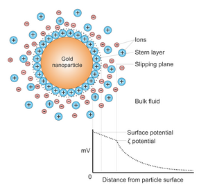
Potential departure as a function of distance from particle surface.
Mostly, gold nanoparticles are produced in a liquid ("liquid chemical methods") by reduction of chloroauric acid (H[AuCl
4 ]).To forbid the particles from aggregating, stabilizing agents are added.Citrate acts both as the reducing amanuensis and colloidal stabilizer.
They tin can be functionalized with diverse organic ligands to create organic-inorganic hybrids with advanced functionality.[17]
Turkevich method [edit]
This simple method was pioneered by J. Turkevich et al. in 1951[105] [106] and refined past G. Frens in the 1970s.[107] [108] It produces modestly monodisperse spherical gold nanoparticles of around ten–20 nm in diameter.Larger particles tin be produced, but at the toll of monodispersity and shape. In this method, hot chloroauric acrid is treated with sodium citrate solution, producing colloidal gold. The Turkevich reaction gain via formation of transient aureate nanowires. These golden nanowires are responsible for the dark appearance of the reaction solution before it turns reddish-red.[109]
Capping agents [edit]
A capping amanuensis is used during nanoparticle synthesis to inhibit particle growth and aggregation. The chemical blocks or reduces reactivity at the periphery of the particle—a good capping agent has a high affinity for the new nuclei.[110] Citrate ions or tannic acid function both as a reducing agent and a capping agent.[111] [112] Less sodium citrate results in larger particles.
Brust-Schiffrin method [edit]
This method was discovered by Brust and Schiffrin in the early 1990s,[113] and can be used to produce gold nanoparticles in organic liquids that are commonly not miscible with water (similar toluene). It involves the reaction of a chlorauric acid solution with tetraoctylammonium bromide (TOAB) solution in toluene and sodium borohydride every bit an anti-coagulant and a reducing agent, respectively.
Here, the gold nanoparticles will be around 5–6 nm.[114] NaBHfour is the reducing amanuensis, and TOAB is both the phase transfer catalyst and the stabilizing agent.
TOAB does not demark to the gold nanoparticles particularly strongly, so the solution volition aggregate gradually over the course of approximately ii weeks. To preclude this, ane can add a stronger binding amanuensis, like a thiol (in particular, alkanethiols), which volition demark to gold, producing a nigh-permanent solution.[115] [116] Alkanethiol protected gold nanoparticles tin exist precipitated and so redissolved. Thiols are better binding agents considering there is a potent affinity for the gold-sulfur bonds that class when the two substances react with each other.[117] Tetra-dodecanthiol is a commonly used strong bounden agent to synthesize smaller particles.[118] Some of the phase transfer agent may remain bound to the purified nanoparticles, this may touch on concrete backdrop such every bit solubility. In gild to remove as much of this agent as possible, the nanoparticles must be further purified by soxhlet extraction.
Perrault method [edit]
This arroyo, discovered by Perrault and Chan in 2009,[119] uses hydroquinone to reduce HAuCl4 in an aqueous solution that contains 15 nm gold nanoparticle seeds. This seed-based method of synthesis is similar to that used in photographic film development, in which silver grains within the film abound through addition of reduced silverish onto their surface. Likewise, gilded nanoparticles can act in conjunction with hydroquinone to catalyze reduction of ionic gilded onto their surface. The presence of a stabilizer such as citrate results in controlled deposition of aureate atoms onto the particles, and growth. Typically, the nanoparticle seeds are produced using the citrate method. The hydroquinone method complements that of Frens,[107] [108] as it extends the range of monodispersed spherical particle sizes that tin be produced. Whereas the Frens method is ideal for particles of 12–20 nm, the hydroquinone method can produce particles of at least 30–300 nm.
Martin method [edit]
This simple method, discovered by Martin and Eah in 2010,[120] generates nearly monodisperse "naked" gold nanoparticles in water. Precisely decision-making the reduction stoichiometry by adjusting the ratio of NaBHfour-NaOH ions to HAuCl4-HCl ions within the "sweet zone," along with heating, enables reproducible diameter tuning betwixt iii–6 nm. The aqueous particles are colloidally stable due to their high accuse from the backlog ions in solution. These particles can be coated with various hydrophilic functionalities, or mixed with hydrophobic ligands for applications in non-polar solvents. In not-polar solvents the nanoparticles remain highly charged, and self-assemble on liquid droplets to form second monolayer films of monodisperse nanoparticles.
Nanotech studies [edit]
Bacillus licheniformis can exist used in synthesis of golden nanocubes with sizes betwixt 10 and 100 nanometres.[121] Gold nanoparticles are usually synthesized at high temperatures in organic solvents or using toxic reagents. The bacteria produce them in much milder conditions.
[edit]
For particles larger than 30 nm, control of particle size with a low polydispersity of spherical gold nanoparticles remains challenging. In order to provide maximum control on the NP structure, Navarro and co-workers used a modified Turkevitch-Frens process using sodium acetylacetonate as the reducing amanuensis and sodium citrate every bit the stabilizer.[122]
Sonolysis [edit]
Another method for the experimental generation of gilded particles is by sonolysis. The first method of this type was invented past Baigent and Müller.[123] This work pioneered the apply of ultrasound to provide the free energy for the processes involved and allowed the creation of gold particles with a diameter of under ten nm. In another method using ultrasound, the reaction of an aqueous solution of HAuClfour with glucose,[124] the reducing agents are hydroxyl radicals and saccharide pyrolysis radicals (forming at the interfacial region between the collapsing cavities and the bulk water) and the morphology obtained is that of nanoribbons with width 30–50 nm and length of several micrometers. These ribbons are very flexible and tin can bend with angles larger than 90°. When glucose is replaced past cyclodextrin (a glucose oligomer), only spherical gold particles are obtained, suggesting that glucose is essential in directing the morphology toward a ribbon.
Block copolymer-mediated method [edit]
An economical, environmentally benign and fast synthesis methodology for golden nanoparticles using cake copolymer has been adult by Sakai et al.[125] In this synthesis methodology, cake copolymer plays the dual office of a reducing agent as well every bit a stabilizing amanuensis. The germination of gold nanoparticles comprises 3 principal steps: reduction of gold salt ion past block copolymers in the solution and germination of gold clusters, adsorption of block copolymers on gilded clusters and farther reduction of gold table salt ions on the surfaces of these golden clusters for the growth of gold particles in steps, and finally its stabilization past block copolymers. Merely this method commonly has a limited-yield (nanoparticle concentration), which does not increase with the increment in the gold salt concentration. Ray et al.[126] improved this synthesis method by enhancing the nanoparticle yield by manyfold at ambient temperature.
See also [edit]
- Colloidal silver
- Gold nanorods
- Gold nanoparticles in chemotherapy
- Nanozymes
References [edit]
- ^ Voliani, Valerio (2020-04-20). Aureate Nanoparticles: An Introduction to Synthesis, Backdrop and Applications. De Gruyter. doi:x.1515/9781501511455. ISBN978-1-5015-1145-5.
- ^ Sapsford KE, Algar WR, Berti Fifty, Gemmill KB, Casey BJ, Oh East, Stewart MH, Medintz IL (March 2013). "Functionalizing nanoparticles with biological molecules: developing chemistries that facilitate nanotechnology". Chemical Reviews. 113 (3): 1904–2074. doi:ten.1021/cr300143v. PMID 23432378.
- ^ Sreekumar, South.; Shah, N.; Mondol, J.; Hewitt, N.; Chakrabarti, Due south. (February 2022). "Broadband arresting mono, composite and hybrid nanofluids for directly absorption solar collector: A comprehensive review". Nano Futures. 103: 504–515. doi:10.1088/2399-1984/ac57f7.
- ^ Gorji, Saleh; Cheong, Kuan Yew (2015). "Au nanoparticles embedded at the interface of Al/4H-SiC Schottky contacts for electric current density enhancement". Applied Physics A. 118: 315–325. doi:10.1007/s00339-014-8733-iv. S2CID 96824985.
- ^ Torres-Torres, D.; Trejo-Valdez, One thousand.; Castañeda, L.; Torres-Torres, C.; Tamayo-Rivera, Fifty.; Fernández-Hernández, R. C.; Reyes-Esqueda, J. A.; Muñoz-Saldaña, J.; Rangel-Rojo, R.; Oliver, A. (2010-08-02). "Inhibition of the two-photon absorption response exhibited past a bilayer TiO2 motion-picture show with embedded Au nanoparticles". Eyes Express. 18 (sixteen): 16406–16417. doi:10.1364/OE.eighteen.016406. ISSN 1094-4087.
- ^ Yang X, Yang M, Pang B, Vara G, Xia Y (October 2015). "Gilt Nanomaterials at Work in Biomedicine". Chemic Reviews. 115 (nineteen): 10410–88. doi:10.1021/acs.chemrev.5b00193. PMID 26293344.
- ^ Mulvaney P (2003). The dazzler and elegance of Nanocrystals: How invisibly small-scale particles will colour and shape our time to come (Report). University of Melbourne. Archived from the original on 2004-x-28.
- ^ Rao CN, Kulkarni GU, Thomas PJ, Edwards PP (2000). "Metallic nanoparticles and their assemblies". Chemic Order Reviews. 29 (1): 27–35. doi:10.1039/A904518J. S2CID 59025862.
- ^ Dreaden EC, Alkilany AM, Huang X, Potato CJ, El-Sayed MA (April 2012). "The gilded age: aureate nanoparticles for biomedicine". Chemical Society Reviews. 41 (7): 2740–79. doi:10.1039/c1cs15237h. PMC5876014. PMID 22109657.
- ^ Zeng Southward, Yong KT, Roy I, Dinh XQ, Yu X, Luan F (2011). "A review on functionalized golden nanoparticles for biosensing applications" (PDF). Plasmonics. half-dozen (3): 491–506. doi:10.1007/s11468-011-9228-1. S2CID 34796473.
- ^ a b Sharma V, Park K, Srinivasarao M (2009). "Colloidal dispersion of gold nanorods: Historical background, optical properties, seed-mediated synthesis, shape separation and self-assembly". Materials Science and Engineering science: R: Reports. 65 (1–3): 1–38. doi:10.1016/j.mser.2009.02.002.
- ^ "The Lycurgus Loving cup". British Museum . Retrieved 2015-12-04 .
- ^ Freestone I, Meeks N, Sax Chiliad, Higgitt C (2007). "The Lycurgus Cup — A Roman nanotechnology". Gold Message. 40 (4): 270–277. doi:10.1007/BF03215599.
- ^ Antonii F (1618). Panacea aurea sive Tractatus duo de ipsius auro potabili. Ex Bibliopolio Frobeniano.
- ^ Culpeper N (1657). Mr. Culpepper'due south Treatise of aurum potabile Being a description of the three-fold earth, viz. elementary angelic intellectual containing the noesis necessary to the study of hermetick philosophy. Faithfully written by him in his life-time, and since his death, published by his wife. London.
- ^ Kunckel von Löwenstern J (1678). Utiles observationes sive animadversiones de salibus fixis et volatilibus, auro et argento potabili (etc.). Austria: Wilson.
- ^ a b Reddy VR (July 2006). "Gold nanoparticles: synthesis and applications". Synlett. 2006 (eleven): 1791–2. doi:10.1055/s-2006-944219.
- ^ a b Faraday M (January 1857). "The Bakerian Lecture: Experimental Relations of Aureate (and Other Metals) to Light". Philosophical Transactions of the Royal Society of London. 147: 145–181. Bibcode:1857RSPT..147..145F. doi:10.1098/rstl.1857.0011.
- ^ "Michael Faraday'southward gilt colloids | The Purple Institution: Science Lives Here". www.rigb.org . Retrieved 2015-12-04 .
- ^ Gay-Lussac (1832). "Ueber den Cassius'schen Goldpurpur". Annalen der Physik. 101 (viii): 629–630. Bibcode:1832AnP...101..629G. doi:10.1002/andp.18321010809.
- ^ Berzelius JJ (1831). "Ueber den Cassius' schen Goldpurpur". Annalen der Physik. 98 (6): 306–308. Bibcode:1831AnP....98..306B. doi:10.1002/andp.18310980613.
- ^ Zsigmondy R (December eleven, 1926). "Properties of colloids" (PDF). Nobel Foundation. Retrieved 2009-01-23 .
- ^ Zeng Southward, Yu X, Law West, Zhang Y, Hu R, Dinh X, H o H, Yong Thousand (2013). "Size dependence of Au NP-enhanced surface plasmon resonance based on differential phase measurement". Sensors and Actuators B: Chemic. 176: 1128–1133. doi:10.1016/j.snb.2012.09.073.
- ^ Anderson ML, Morris CA, Stroud RM, Merzbacher CI, Rolison DR (1999-02-01). "Colloidal Gold Aerogels: Training, Backdrop, and Label". Langmuir. 15 (three): 674–681. doi:10.1021/la980784i.
- ^ a b Link S, El-Sayed MA (1999-05-01). "Size and Temperature Dependence of the Plasmon Assimilation of Colloidal Gilded Nanoparticles". The Journal of Physical Chemistry B. 103 (21): 4212–4217. CiteSeerXx.1.1.596.6328. doi:10.1021/jp984796o.
- ^ a b Ghosh SK, Nath S, Kundu South, Esumi Thou, Pal T (2004-09-01). "Solvent and Ligand Effects on the Localized Surface Plasmon Resonance (LSPR) of Golden Colloids". The Journal of Physical Chemistry B. 108 (37): 13963–13971. doi:ten.1021/jp047021q.
- ^ a b Underwood S, Mulvaney P (1994-10-01). "Effect of the Solution Refractive Index on the Color of Aureate Colloids". Langmuir. 10 (ten): 3427–3430. doi:10.1021/la00022a011.
- ^ Xing S, Tan LH, Yang One thousand, Pan M, Lv Y, Tang Q, Yang Y, Chen H (2009-05-12). "Highly controlled core/shell structures: tunable conductive polymer shells on gold nanoparticles and nanochains". Journal of Materials Chemical science. 19 (20): 3286. doi:10.1039/b900993k. S2CID 96293198.
- ^ Ghosh SK, Pal T (November 2007). "Interparticle coupling result on the surface plasmon resonance of gold nanoparticles: from theory to applications". Chemical Reviews. 107 (xi): 4797–862. doi:ten.1021/cr0680282. PMID 17999554. S2CID 46326525.
- ^ Horisberger M, Rosset J (April 1977). "Colloidal aureate, a useful marker for manual and scanning electron microscopy". The Journal of Histochemistry and Cytochemistry. 25 (four): 295–305. doi:ten.1177/25.4.323352. PMID 323352.
- ^ Electron microscopy: principles and techniques for biologists (2nd ed.). Jones and Bartlett. Oct 1998. ISBN978-0-7637-0192-5.
- ^ Hunter EE (September 1993). Practical electron microscopy : a beginner'southward illustrated guide (2nd ed.). Cambridge University Press. ISBN978-0-521-38539-eight.
- ^ Electron microscopy: methods and protocols. Methods in Molecular Biology (2nd ed.). Humana Press. Feb 2007. ISBN978-i-58829-573-half dozen.
- ^ Romano EL, Romano K (1977). "Staphylococcal protein a spring to colloidal gilded: A useful reagent to characterization antigen-antibody sites in electron microscopy". Immunochemistry. 14 (9–10): 711–715. doi:10.1016/0019-2791(77)90146-10.
- ^ Fetni R, Drouin R, Lemieux N, Messier PE, Richer CL (December 1991). "Simultaneous visualization of chromosome bands and hybridization signal using colloidal-gilded labeling in electron microscopy". Proceedings of the National University of Sciences of the U.s. of America. 88 (23): 10916–20. Bibcode:1991PNAS...8810916F. doi:10.1073/pnas.88.23.10916. PMC53043. PMID 1961763.
- ^ Kasamatsu H, Lin W, Edens J, Revel JP (July 1983). "Visualization of antigens fastened to cytoskeletal framework in animate being cells: colocalization of simian virus 40 Vp1 polypeptide and actin in TC7 cells". Proceedings of the National Academy of Sciences of the Usa. 80 (14): 4339–43. Bibcode:1983PNAS...80.4339K. doi:10.1073/pnas.80.xiv.4339. PMC384033. PMID 6308616.
Double labeling with colloidal gilded particles of unlike sizes
- ^ Grobelny J, DelRio FW, Pradeep N, Kim DI, Hackley VA, Melt RF (2011). "Size measurement of nanoparticles using atomic force microscopy". In McNeil SE (ed.). Characterization of nanoparticles intended for drug delivery. Humana Press. pp. 71–82. ISBN978-i-60327-198-1.
- ^ Han Thousand, Ghosh P, Rotello VM (Feb 2007). "Functionalized gold nanoparticles for drug delivery". Nanomedicine. 2 (1): 113–23. doi:ten.2217/17435889.2.i.113. PMID 17716197.
- ^ Han G, Ghosh P, Rotello VM (2007). "Multi-functional gilded nanoparticles for drug delivery". Bio-Applications of Nanoparticles. Advances in Experimental Medicine and Biology. Vol. 620. pp. 48–56. doi:ten.1007/978-0-387-76713-0_4. ISBN978-0-387-76712-3. PMID 18217334.
- ^ Langer R (February 2000). "Biomaterials in drug delivery and tissue technology: i laboratory's experience". Accounts of Chemical Inquiry. 33 (two): 94–101. doi:x.1021/ar9800993. PMID 10673317.
- ^ Gibson JD, Khanal BP, Zubarev ER (September 2007). "Paclitaxel-functionalized gilded nanoparticles". Journal of the American Chemical Guild. 129 (37): 11653–61. doi:ten.1021/ja075181k. PMID 17718495. S2CID 12034022.
- ^ Qian X, Peng XH, Ansari Practice, Yin-Goen Q, Chen GZ, Shin DM, Yang L, Young AN, Wang Medico, Nie Due south (January 2008). "In vivo tumor targeting and spectroscopic detection with surface-enhanced Raman nanoparticle tags". Nature Biotechnology. 26 (one): 83–90. doi:10.1038/nbt1377. PMID 18157119. S2CID 15309464.
- ^ Sajjadi AY, Suratkar AA, Mitra KK, Grace MS (2012). "Short-Pulse Laser-Based System for Detection of Tumors: Assistants of Gold Nanoparticles Enhances Contrast". Journal of Nanotechnology in Technology and Medicine. 3 (two): 021002. doi:10.1115/one.4007245.
- ^ Giljohann DA, Seferos DS, Prigodich AE, Patel PC, Mirkin CA. Gene regulation with polyvalent siRNA-nanoparticle conjugates. J Am Chem Soc 2009;131:2072–2073.
- ^ Mackey MA, Ali MR, Austin LA, Nearly RD, El-Sayed MA (Feb 2014). "The most constructive gold nanorod size for plasmonic photothermal therapy: theory and in vitro experiments". The Journal of Physical Chemistry B. 118 (v): 1319–26. doi:10.1021/jp409298f. PMC3983380. PMID 24433049.
- ^ Niidome T, Yamagata Grand, Okamoto Y, Akiyama Y, Takahashi H, Kawano T, Katayama Y, Niidome Y (September 2006). "PEG-modified gold nanorods with a stealth graphic symbol for in vivo applications". Journal of Controlled Release. 114 (three): 343–7. doi:x.1016/j.jconrel.2006.06.017. PMID 16876898.
- ^ Cassano, Domenico; Pocoví-Martínez, Salvador; Voliani, Valerio (2018-01-17). "Ultrasmall-in-Nano Approach: Enabling the Translation of Metal Nanomaterials to Clinics". Bioconjugate Chemistry. 29 (ane): four–16. doi:x.1021/acs.bioconjchem.7b00664. ISSN 1043-1802. PMID 29186662.
- ^ Vlamidis, Ylea; Voliani, Valerio (2018-x-08). "Bringing Again Noble Metal Nanoparticles to the Forefront of Cancer Therapy". Frontiers in Bioengineering and Biotechnology. half-dozen: 143. doi:x.3389/fbioe.2018.00143. ISSN 2296-4185. PMC6186777. PMID 30349817.
- ^ Cassano, Domenico; Santi, Melissa; D'Autilia, Francesca; Mapanao, Ana Katrina; Luin, Stefano; Voliani, Valerio (2019). "Photothermal outcome past NIR-responsive excretable ultrasmall-in-nano architectures". Materials Horizons. 6 (3): 531–537. doi:ten.1039/C9MH00096H. ISSN 2051-6347.
- ^ Hainfeld JF, Slatkin DN, Smilowitz HM (September 2004). "The employ of gilt nanoparticles to enhance radiotherapy in mice". Physics in Medicine and Biology. 49 (18): N309–15. doi:10.1088/0031-9155/49/18/N03. PMID 15509078. S2CID 28457097.
- ^ McMahon SJ, Hyland WB, Muir MF, Coulter JA, Jain South, Butterworth KT, Schettino Grand, Dickson GR, Hounsell AR, O'Sullivan JM, Prise KM, Hirst DG, Currell FJ (2011). "Biological consequences of nanoscale energy degradation nigh irradiated heavy cantlet nanoparticles". Scientific Reports. 1: 18. Bibcode:2011NatSR...1E..18M. doi:10.1038/srep00018. PMC3216506. PMID 22355537.
- ^ Zhang Z, Chen Z, Wang Due south, Qu C, Chen L (May 2014). "On-site visual detection of hydrogen sulfide in air based on enhancing the stability of aureate nanoparticles". ACS Practical Materials & Interfaces. 6 (ix): 6300–7. doi:10.1021/am500564w. PMID 24754960.
- ^ a b c d east Xu South (2010). "Gilded nanoparticle-based biosensors". Aureate Bulletin. 43: 29–41. doi:10.1007/BF03214964.
- ^ Wang J, Polsky R, Xu D (2001). "Silver-Enhanced Colloidal Gold Electrochemical Stripping Detection of Dna Hybridization". Langmuir. 17 (xix): 5739. doi:10.1021/la011002f.
- ^ Wang J, Xu D, Polsky R (Apr 2002). "Magnetically-induced solid-country electrochemical detection of DNA hybridization". Journal of the American Chemic Society. 124 (xvi): 4208–9. doi:10.1021/ja0255709. PMID 11960439.
- ^ Daniel MC, Astruc D (Jan 2004). "Gold nanoparticles: assembly, supramolecular chemistry, quantum-size-related properties, and applications toward biological science, catalysis, and nanotechnology". Chemic Reviews. 104 (1): 293–346. doi:10.1021/cr030698+. PMID 14719978. S2CID 29293663.
- ^ Hu M, Chen J, Li ZY, Au L, Hartland GV, Li X, Marquez M, Xia Y (November 2006). "Gold nanostructures: technology their plasmonic backdrop for biomedical applications". Chemic Society Reviews. 35 (11): 1084–94. doi:10.1039/b517615h. PMID 17057837. S2CID 2259806.
- ^ Link S, El-Sayed MA (1996). "Spectral Properties and Relaxation Dynamics of Surface Plasmon Electronic Oscillations in Gold and Argent Nanodots and Nanorods". J. Phys. Chem. B. 103 (40): 8410. doi:10.1021/jp9917648. S2CID 40012876.
- ^ Mulvaney, P. (1996). "Surface Plasmon Spectroscopy of Nanosized Metal Particles". Langmuir. 12 (3): 788. doi:x.1021/la9502711.
- ^ Lin HY, Chen CT, Chen YC (October 2006). "Detection of phosphopeptides by localized surface plasma resonance of titania-coated gilt nanoparticles immobilized on glass substrates". Analytical Chemistry. 78 (19): 6873–8. doi:10.1021/ac060833t. PMID 17007509. S2CID 13373307.
- ^ He L, Musick MD, Nicewarner SR, Salinas FG (2000). "Colloidal Au-Enhanced Surface Plasmon Resonance for Ultrasensitive Detection of DNA Hybridization". Periodical of the American Chemical Society. 122 (38): 9071. doi:ten.1021/ja001215b.
- ^ Okamoto T, Yamaguchi I, Kobayashi T (2000). "Local plasmon sensor with gold colloid monolayers deposited upon glass substrates". Opt Lett. 25 (6): 372–four. Bibcode:2000OptL...25..372O. doi:10.1364/OL.25.000372. PMID 18059883.
- ^ Dark-brown KR, Fox P, Natan MJ (1996). "Morphology-Dependent Electrochemistry of Cytochromecat Au Colloid-Modified SnO2Electrodes". Journal of the American Chemical Society. 118 (5): 1154. doi:10.1021/ja952951w.
- ^ Xiao Y, Patolsky F, Katz E, Hainfeld JF, Willner I (March 2003). ""Plugging into Enzymes": nanowiring of redox enzymes past a gold nanoparticle". Scientific discipline. 299 (5614): 1877–81. Bibcode:2003Sci...299.1877X. doi:10.1126/science.1080664. PMID 12649477. S2CID 40388898.
- ^ Gole A, Dash C, Ramakrishnan V, Sainkar SR, Mandale AB, Rao M, Sastry M (2001). "Pepsin−Gold Colloid Conjugates: Training, Characterization, and Enzymatic Activeness". Langmuir. 17 (5): 1674. doi:10.1021/la001164w.
- ^ Gole A, Vyas Due south, Phadtare S, Lachke A, Sastry Thousand (2002). "Studies on the germination of bioconjugates of Endoglucanase with colloidal gold". Colloids and Surfaces B: Biointerfaces. 25 (2): 129. doi:10.1016/s0927-7765(01)00301-0.
- ^ Valden M, Lai 10, Goodman DW (September 1998). "Onset of catalytic activity of gold clusters on titania with the appearance of nonmetallic properties". Science. 281 (5383): 1647–50. Bibcode:1998Sci...281.1647V. doi:10.1126/science.281.5383.1647. PMID 9733505. S2CID 21287894.
- ^ Lou Y, Maye MM, Han L, Zhong CJ (2001). "Golden–platinum alloy nanoparticle associates as catalyst for methanol electrooxidation". Chemical Communications. 2001 (5): 473. doi:10.1039/b008669j.
- ^ Turner Grand, Golovko VB, Vaughan OP, Abdulkin P, Berenguer-Murcia A, Tikhov MS, Johnson BF, Lambert RM (August 2008). "Selective oxidation with dioxygen by golden nanoparticle catalysts derived from 55-atom clusters". Nature. 454 (7207): 981–iii. Bibcode:2008Natur.454..981T. doi:10.1038/nature07194. PMID 18719586. S2CID 4355469.
- ^ Marradi M, Chiodo F, García I, Penadés S (2013). "Glyconanoparticles as multifunctional and multimodal carbohydrate systems". Chem. Soc. Rev. 42 (11): 4728–45. doi:x.1039/C2CS35420A. PMID 23288339.
- ^ Chiodo F, Marradi M, Tefsen B, Snippe H, van Dice I, Penadés South (2013). "Loftier sensitive detection of carbohydrate binding proteins in an ELISA-solid phase assay based on multivalent glyconanoparticles". PLOS 1. 8 (eight): e73027. Bibcode:2013PLoSO...873027C. doi:ten.1371/journal.pone.0073027. PMC3754922. PMID 24014084.
- ^ Mueggenburg KE, Lin XM, Goldsmith RH, Jaeger HM (September 2007). "Rubberband membranes of close-packed nanoparticle arrays". Nature Materials. 6 (nine): 656–60. Bibcode:2007NatMa...6..656M. doi:ten.1038/nmat1965. PMID 17643104. S2CID 444592.
- ^ He J, Kanjanaboos P, Frazer NL, Weis A, Lin XM, Jaeger HM (July 2010). "Fabrication and mechanical properties of large-scale freestanding nanoparticle membranes". Small. six (xiii): 1449–56. doi:10.1002/smll.201000114. PMID 20521265. S2CID 206491859.
- ^ Wang Y, Kanjanaboos P, Barry Due east, McBride Southward, Lin XM, Jaeger HM (February 2014). "Fracture and failure of nanoparticle monolayers and multilayers". Nano Letters. 14 (2): 826–30. Bibcode:2014NanoL..xiv..826W. doi:10.1021/nl404185b. PMID 24467462. S2CID 207673690.
- ^ Wang Y, Liao J, McBride SP, Efrati E, Lin XM, Jaeger HM (October 2015). "Strong Resistance to Angle Observed for Nanoparticle Membranes". Nano Letters. xv (ten): 6732–7. Bibcode:2015NanoL..15.6732W. doi:10.1021/acs.nanolett.5b02587. PMID 26313627. S2CID 29849022.
- ^ Griesemer SD, You SS, Kanjanaboos P, Calabro Thou, Jaeger HM, Rice SA, Lin B (May 2017). "The part of ligands in the mechanical properties of Langmuir nanoparticle films". Soft Matter. xiii (17): 3125–3133. Bibcode:2017SMat...13.3125G. doi:10.1039/c7sm00319f. PMID 28397901.
- ^ a b c Sperling RA, Parak WJ (March 2010). "Surface modification, functionalization and bioconjugation of colloidal inorganic nanoparticles". Philosophical Transactions. Series A, Mathematical, Physical, and Technology Sciences. 368 (1915): 1333–83. Bibcode:2010RSPTA.368.1333S. doi:ten.1098/rsta.2009.0273. PMID 20156828.
- ^ Tauran Y, Brioude A, Coleman AW, Rhimi K, Kim B (August 2013). "Molecular recognition by gold, silvery and copper nanoparticles". World Journal of Biological Chemistry. 4 (3): 35–63. doi:10.4331/wjbc.v4.i3.35. PMC3746278. PMID 23977421.
- ^ Taguchi T, Isozaki One thousand, Miki K (December 2012). "Enhanced catalytic activity of self-assembled-monolayer-capped gold nanoparticles". Avant-garde Materials. 24 (48): 6462–seven. doi:ten.1002/adma.201202979. PMID 22968900. S2CID 205247206.
- ^ Heinecke CL, Ni TW, Malola Due south, Mäkinen V, Wong OA, Häkkinen H, Ackerson CJ (August 2012). "Structural and theoretical basis for ligand commutation on thiolate monolayer protected gold nanoclusters". Journal of the American Chemical Society. 134 (32): 13316–22. doi:10.1021/ja3032339. PMC4624284. PMID 22816317.
- ^ Perumal S, Hofmann A, Scholz Northward, Rühl E, Graf C (April 2011). "Kinetics study of the bounden of multivalent ligands on size-selected gold nanoparticles". Langmuir. 27 (8): 4456–64. doi:ten.1021/la105134m. PMID 21413796.
- ^ McMahon JM, Emory SR (Jan 2007). "Stage transfer of big gilded nanoparticles to organic solvents with increased stability". Langmuir. 23 (three): 1414–8. doi:10.1021/la0617560. PMID 17241067.
- ^ Tyo EC, Vajda S (July 2015). "Catalysis by clusters with precise numbers of atoms". Nature Nanotechnology. ten (7): 577–88. Bibcode:2015NatNa..10..577T. doi:ten.1038/nnano.2015.140. PMID 26139144.
- ^ Niu Z, Li Y (2014-01-14). "Removal and Utilization of Capping Agents in Nanocatalysis". Chemistry of Materials. 26 (1): 72–83. doi:x.1021/cm4022479.
- ^ Häkkinen H, Walter M, Grönbeck H (May 2006). "Divide and protect: capping aureate nanoclusters with molecular gold-thiolate rings". The Journal of Physical Chemistry B. 110 (20): 9927–31. doi:10.1021/jp0619787. PMID 16706449.
- ^ Reimers JR, Ford MJ, Halder A, Ulstrup J, Hush NS (March 2016). "Gold surfaces and nanoparticles are protected past Au(0)-thiyl species and are destroyed when Au(I)-thiolates grade". Proceedings of the National Academy of Sciences of the U.s.a. of America. 113 (eleven): E1424–33. Bibcode:2016PNAS..113E1424R. doi:x.1073/pnas.1600472113. PMC4801306. PMID 26929334.
- ^ Park JW, Shumaker-Parry JS (February 2014). "Structural study of citrate layers on gold nanoparticles: role of intermolecular interactions in stabilizing nanoparticles". Periodical of the American Chemical Order. 136 (5): 1907–21. doi:10.1021/ja4097384. PMID 24422457.
- ^ a b c Alkilany AM, Murphy CJ (September 2010). "Toxicity and cellular uptake of gilded nanoparticles: what we have learned and so far?". Journal of Nanoparticle Inquiry. 12 (7): 2313–2333. Bibcode:2010JNR....12.2313A. doi:ten.1007/s11051-010-9911-8. PMC2988217. PMID 21170131.
- ^ Perala SR, Kumar Due south (Baronial 2013). "On the mechanism of metal nanoparticle synthesis in the Brust-Schiffrin method". Langmuir. 29 (31): 9863–73. doi:10.1021/la401604q. PMID 23848382.
- ^ a b c Alkilany AM, Nagaria PK, Hexel CR, Shaw TJ, Murphy CJ, Wyatt Physician (March 2009). "Cellular uptake and cytotoxicity of gold nanorods: molecular origin of cytotoxicity and surface effects". Small. five (6): 701–8. doi:10.1002/smll.200801546. PMID 19226599.
- ^ Takahashi H, Niidome Y, Niidome T, Kaneko Grand, Kawasaki H, Yamada S (January 2006). "Modification of gold nanorods using phosphatidylcholine to reduce cytotoxicity". Langmuir. 22 (one): ii–5. doi:10.1021/la0520029. PMID 16378388.
- ^ a b c Goodman CM, McCusker CD, Yilmaz T, Rotello VM (June 2004). "Toxicity of gilt nanoparticles functionalized with cationic and anionic side chains". Bioconjugate Chemistry. 15 (4): 897–900. doi:10.1021/bc049951i. PMID 15264879.
- ^ Connor EE, Mwamuka J, Gole A, Murphy CJ, Wyatt Md (March 2005). "Gilt nanoparticles are taken upward past human being cells merely do non crusade acute cytotoxicity". Small. 1 (3): 325–vii. doi:10.1002/smll.200400093. PMID 17193451.
- ^ Dobrovolskaia MA, Patri AK, Zheng J, Clogston JD, Ayub N, Aggarwal P, Neun BW, Hall JB, McNeil SE (June 2009). "Interaction of colloidal gilded nanoparticles with human blood: effects on particle size and analysis of plasma protein binding profiles". Nanomedicine. five (2): 106–17. doi:x.1016/j.nano.2008.08.001. PMC3683956. PMID 19071065.
- ^ Chen YS, Hung YC, Liau I, Huang GS (May 2009). "Cess of the In Vivo Toxicity of Golden Nanoparticles". Nanoscale Enquiry Messages. 4 (viii): 858–864. Bibcode:2009NRL.....iv..858C. doi:10.1007/s11671-009-9334-half dozen. PMC2894102. PMID 20596373.
- ^ Cho WS, Cho Thousand, Jeong J, Choi M, Cho HY, Han BS, Kim SH, Kim HO, Lim YT, Chung BH, Jeong J (April 2009). "Acute toxicity and pharmacokinetics of 13 nm-sized PEG-coated gilded nanoparticles". Toxicology and Applied Pharmacology. 236 (1): xvi–24. doi:10.1016/j.taap.2008.12.023. PMID 19162059.
- ^ Gref R, Couvreur P, Barratt G, Mysiakine East (November 2003). "Surface-engineered nanoparticles for multiple ligand coupling". Biomaterials. 24 (24): 4529–37. doi:x.1016/s0142-9612(03)00348-ten. PMID 12922162.
- ^ Boisselier E, Astruc D (June 2009). "Gold nanoparticles in nanomedicine: preparations, imaging, diagnostics, therapies and toxicity". Chemical Social club Reviews. 38 (vi): 1759–82. doi:10.1039/b806051g. PMID 19587967.
- ^ Tandon A, Sharma A, Rodier JT, Klibanov AM, Rieger FG, Mohan RR (June 2013). "BMP7 gene transfer via gilt nanoparticles into stroma inhibits corneal fibrosis in vivo". PLOS ONE. 8 (6): e66434. Bibcode:2013PLoSO...866434T. doi:10.1371/journal.pone.0066434. PMC3682981. PMID 23799103.
- ^ Gratton SE, Pohlhaus PD, Lee J, Guo J, Cho MJ, Desimone JM (August 2007). "Nanofabricated particles for engineered drug therapies: a preliminary biodistribution report of PRINT nanoparticles". Periodical of Controlled Release. 121 (1–ii): 10–8. doi:10.1016/j.jconrel.2007.05.027. PMC1994820. PMID 17643544.
- ^ a b c De Jong WH, Hagens WI, Krystek P, Burger MC, Sips AJ, Geertsma RE (April 2008). "Particle size-dependent organ distribution of golden nanoparticles after intravenous administration". Biomaterials. 29 (12): 1912–9. doi:10.1016/j.biomaterials.2007.12.037. PMID 18242692.
- ^ Cassano, Domenico; Mapanao, Ana-Katrina; Summa, Maria; Vlamidis, Ylea; Giannone, Giulia; Santi, Melissa; Guzzolino, Elena; Pitto, Letizia; Poliseno, Laura; Bertorelli, Rosalia; Voliani, Valerio (2019-10-21). "Biosafety and Biokinetics of Noble Metals: The Impact of Their Chemic Nature". ACS Applied Bio Materials. 2 (ten): 4464–4470. doi:10.1021/acsabm.9b00630. ISSN 2576-6422.
- ^ Cassano, Domenico; Summa, Maria; Pocoví-Martínez, Salvador; Mapanao, Ana-Katrina; Catelani, Tiziano; Bertorelli, Rosalia; Voliani, Valerio (Feb 2019). "Biodegradable Ultrasmall-in-Nano Gilded Architectures: Mid-Menstruation In Vivo Distribution and Excretion Assessment". Particle & Particle Systems Characterization. 36 (2): 1800464. doi:10.1002/ppsc.201800464.
- ^ Turkevich J, Stevenson PC, Hillier J (1951). "A report of the nucleation and growth processes in the synthesis of colloidal gold". Discuss. Faraday Soc. 11: 55–75. doi:ten.1039/df9511100055. S2CID 97664009.
- ^ Kimling J, Maier M, Okenve B, Kotaidis V, Ballot H, Plech A (August 2006). "Turkevich method for gilded nanoparticle synthesis revisited". The Journal of Concrete Chemistry B. 110 (32): 15700–vii. doi:ten.1021/jp061667w. PMID 16898714. S2CID 11729630.
- ^ a b Frens, Grand. (1972). "Particle size and sol stability in metal colloids". Colloid & Polymer Scientific discipline. 250 (7): 736–741. doi:10.1007/bf01498565. S2CID 92726968.
- ^ a b Frens, G. (1973). "Controlled nucleation for the regulation of the particle size in monodisperse gold suspensions". Nature. 241 (105): 20–22. Bibcode:1973NPhS..241...20F. doi:10.1038/physci241020a0.
- ^ Pong BK, Elim Hi, Chong JX, Trout BL, Lee JY (2007). "New Insights on the Nanoparticle Growth Mechanism in the Citrate Reduction of Gold(III) Salt: Formation of the Au Nanowire Intermediate and Its Nonlinear Optical Properties". J. Phys. Chem. C. 111 (17): 6281–6287. doi:10.1021/jp068666o.
- ^ Niu Z, Li Y (2014). "Removal and Utilization of Capping Agents in Nanocatalysis". Chemical science of Materials. 26: 72–83. doi:10.1021/cm4022479.
- ^ Fang Y, Tan J, Lan T, Foo SG, Pyun DG, Lim South, Kim D (2018). "Universal 1‐pot, one‐step synthesis of cadre–shell nanocomposites with self‐assembled tannic acrid shell and their antibacterial and catalytic activities". Journal of Applied Polymer Science. 135 (6): 45829. doi:10.1002/app.45829.
- ^ Fang Y, Tan J, Choi H, Lim S, Kim D (2018). "Highly sensitive naked heart detection of Iron (III) and H2O2 using poly-(tannic acrid) (PTA) coated Au nanocomposite". Sensors and Actuators B: Chemical. 259: 155–161. doi:10.1016/j.snb.2017.12.031.
- ^ Brust M, Walker Chiliad, Bethell D, Schiffrin DJ, Whyman R (1994). "Synthesis of Thiol-derivatised Golden Nanoparticles in a Two-phase Liquid-Liquid System". Chem. Commun. (seven): 801–802. doi:10.1039/C39940000801.
- ^ Manna A, Chen P, Akiyama H, Wei T, Tamada K, Knoll West (2003). "Optimized Photoisomerization on Gilded Nanoparticles Capped by Unsymmetrical Azobenzene Disulfides". Chemistry of Materials. 15 (i): 20–28. doi:10.1021/cm0207696.
- ^ Gao J, Huang X, Liu H, Zan F, Ren J (March 2012). "Colloidal stability of aureate nanoparticles modified with thiol compounds: bioconjugation and application in cancer cell imaging". Langmuir. 28 (9): 4464–71. doi:10.1021/la204289k. PMID 22276658.
- ^ Bekalé, Laurent, Saïd Barazzouk, and Surat Hotchandani. "Beneficial Office of Gold Nanoparticles equally Photoprotector of Magnesium Tetraphenylporphyrin." SpringerReference (northward.d.): due north. pag. Web. 14 Nov. 2016.
- ^ Templeton AC, Wuelfing WP, Murray RW (January 2000). "Monolayer-protected cluster molecules". Accounts of Chemical Research. 33 (1): 27–36. CiteSeerX10.one.1.501.2383. doi:10.1021/ar9602664. PMID 10639073.
- ^ Louis C (2017). "Chemical preparation of gold nanoparticles on surfaces". In Louis C, Pluchery O (eds.). Gilt nanoparticles for physics, chemistry and biological science (2nd ed.). Hackensack (Northward.J.); London: World Scientific. p. 155. ISBN978-1-78634-124-2.
- ^ Perrault SD, Chan WC (December 2009). "Synthesis and surface modification of highly monodispersed, spherical gilt nanoparticles of fifty-200 nm". Journal of the American Chemic Society. 131 (47): 17042–three. doi:10.1021/ja907069u. PMID 19891442.
- ^ Martin MN, Basham JI, Chando P, Eah SK (May 2010). "Charged gold nanoparticles in non-polar solvents: ten-min synthesis and 2D cocky-assembly". Langmuir. 26 (10): 7410–7. doi:x.1021/la100591h. PMID 20392108. A 3-min sit-in video for the Martin synthesis method is available at YouTube
- ^ Kalishwaralal K, Deepak Five, Ram Kumar Pandian Southward, Gurunathan South (November 2009). "Biological synthesis of golden nanocubes from Bacillus licheniformis". Bioresource Technology. 100 (21): 5356–8. doi:10.1016/j.biortech.2009.05.051. PMID 19574037.
- ^ Navarro JR, Lerouge F, Cepraga C, Micouin G, Favier A, Chateau D, Charreyre MT, Lanoë PH, Monnereau C, Chaput F, Marotte S, Leverrier Y, Marvel J, Kamada K, Andraud C, Baldeck PL, Parola S (November 2013). "Nanocarriers with ultrahigh chromophore loading for fluorescence bio-imaging and photodynamic therapy". Biomaterials. 34 (33): 8344–51. doi:10.1016/j.biomaterials.2013.07.032. PMID 23915950.
- ^ Baigent CL, Müller G (1980). "A colloidal gold prepared using ultrasonics". Experientia. 36 (four): 472–473. doi:ten.1007/BF01975154. S2CID 32998274.
- ^ Zhang J, Du J, Han B, Liu Z, Jiang T, Zhang Z (February 2006). "Sonochemical formation of unmarried-crystalline gold nanobelts". Angewandte Chemie. 45 (seven): 1116–9. doi:10.1002/ange.200503762. PMID 16389606.
- ^ Sakai T, Alexandridis P (April 2005). "Mechanism of golden metal ion reduction, nanoparticle growth and size command in aqueous amphiphilic cake copolymer solutions at ambient atmospheric condition". The Periodical of Physical Chemistry B. 109 (16): 7766–77. doi:x.1021/jp046221z. PMID 16851902.
- ^ Ray D, Aswal VK, Kohlbrecher J (March 2011). "Synthesis and Characterization of High Concentration Block Copolymer-Mediated Gold Nanoparticles". Langmuir. 27 (7): 4048–56. doi:x.1021/la2001706. PMID 21366279.
Further reading [edit]
- Boisselier E, Astruc D (June 2009). "Golden nanoparticles in nanomedicine: preparations, imaging, diagnostics, therapies and toxicity". Chemical Club Reviews. 38 (half dozen): 1759–82. doi:10.1039/b806051g. PMID 19587967.
External links [edit]
- Moriarty, Philip. "Au – Gold Nanoparticle". Sixty Symbols. Brady Haran for the University of Nottingham.
- Point-by-point methods for citrate synthesis and hydroquinone synthesis of golden nanoparticles are available here.
Source: https://en.wikipedia.org/wiki/Colloidal_gold
0 Response to "Gold Injected Into Your Cells Gold Used as Art"
Enregistrer un commentaire